Lateral Wall | Atlas of Human Cardiac Anatomy. Location: The lateral wall is generally considered to include the wall of the right atrium from the ostia of the superior and inferior vena cava anteriorly to the ostium of the right appendage or auricle.
What is the lateral wall of the left ventricle?
The left ventricular lateral wall is supplied by branches of the left anterior descending artery (LAD) and left circumflex artery (LCx). Lateral and posterior walls together form the left ventricular free wall which is a common site for free-wall rupture (FWR) post-MI.
What is the inferior side of the heart?
The inferior tip of the heart, the apex, lies just to the left of the sternum between the junction of the fourth and fifth ribs near their articulation with the costal cartilages. The right side of the heart is deflected anteriorly, and the left side is deflected posteriorly.
What are the lateral walls of the atria called?
The flaplike structures on the lateral walls of the atria are called auricles. The auricles are muscular pouches that increase the blood-holding capacity of the atria. They expand to fill and hold blood until an electrical signal thru the appropriate node triggers contraction of the associated atrium.What are the 4 surfaces of the heart?
The heart has five surfaces: base (posterior), diaphragmatic (inferior), sternocostal (anterior), and left and right pulmonary surfaces.
What does lateral mean in anatomy?
Lateral means to the side of, or away from, the middle of the body. Examples: The ears are lateral to the nose. The arms are lateral to the chest.
What are the lateral leads?
The septum is represented on the ECG by leads V1 and V2, whereas the lateral wall is represented by leads V5, V6, lead I and lead aVL.
What is lateral wall infarction?
A lateral myocardial infarction (MI) is a heart attack or cessation of blood flow to the heart muscle that involves the inferior side of the heart. Inferior MI results from the total occlusion of the left circumflex artery. Lateral MI is characterized by ST elevation on the electrocardiogram (EKG) in leads I and aVL.What artery supplies the lateral wall of the heart?
Circumflex artery, which passes behind the heart between the left atrium and left ventricle and supplies blood to the side (lateral wall) of the left ventricle.
What is the anatomy of the heart?The heart is made up of four chambers: two upper chambers known as the left atrium and right atrium and two lower chambers called the left and right ventricles. It is also made up of four valves: the tricuspid, pulmonary, mitral and aortic valves.
Article first time published onWhat forms the inferior border of the heart?
The inferior border of the heart is formed predominantly by the right ventricle. The left ventricle contributes near the apex. It is the most acutely angled border of the heart and it is roughly horizontal.
What forms the right border of the heart?
The right border of the heart extends just over 1 cm to the right of the sternum, between the 3rd and 6th intercostal cartilages. It is composed mainly of the right atrium. The inferior border runs from the 6th costal cartilage on the right, through the xiphisternal joint, to the 5th intercostal space on the left.
What forms the left border of the heart?
Anatomical terminology The left border of heart (or left margin, or obtuse margin) is shorter than the right border, full, and rounded: it is formed mainly by the left ventricle, but to a slight extent, above, by the left atrium.
Where is upper border of the heart?
The upper border is hidden behind the sternum at the level of the second and third cartilages. The right margin of the heart peeps out behind the right border of the sternum between the right third and sixth cartilages.
What is the superior border of the heart?
The superior border of the heart is a line that connects the inferior border of the second left costal cartilage and the superior border of the third right costal cartilage.
What are the 12 parts of the heart?
- Left atrium and auricle. Left atrium. Left auricle.
- Right atrium and auricle. Right atrium. Right auricle.
- Interventricular septum and septal papillary muscles. Interventricular septum. …
- Right ventricle and papillary muscles. Right ventricle. …
- Left ventricle and papillary muscles. Left ventricle.
What does high lateral mean?
High lateral STEMI is associated with a pattern of ST elevation caused by acute occlusion of the first diagonal branch of the left anterior descending coronary artery (LAD-D1).
How do you read a myocardial infarction ECG?
- ST segment elevation in the anterior leads (V3 and V4) at the J point and sometimes in the septal or lateral leads, depending on the extent of the MI. …
- Reciprocal ST segment depression in the inferior leads (II, III and aVF).
What causes lateral ischemia?
Causes of myocardial ischemia Myocardial ischemia occurs when blood flow to your heart is reduced, preventing the heart muscle from receiving enough oxygen. The reduced blood flow is usually the result of a partial or complete blockage of your heart’s arteries (coronary arteries).
Where are your lateral?
A lateral orientation is a position away from the midline of the body. For instance, the arms are lateral to the chest, and the ears are lateral to the head. A medial orientation is a position toward the midline of the body.
What is lateral left?
Depending on the side of the body on which the patient is being operated, the patient will lie on their left or right side. In the left lateral position, the patient lies on the left side of their body for a surgical procedure on their right side.
What is lateral position?
Definition. Lateral position. The lateral position is described as side‐lying with pillows strategically placed along the patient’s back, and possibly buttocks, and a pillow placed between the patient’s flexed legs to prevent adduction and internal rotation of the hip.
Which artery is the most common to have blockage?
Although blockages can occur in other arteries leading to the heart, the LAD artery is where most blockages occur.
What part of the heart does the RCA supply?
Right coronary artery (RCA). The right coronary artery supplies blood to the right ventricle, the right atrium, and the SA (sinoatrial) and AV (atrioventricular) nodes, which regulate the heart rhythm.
What are the 3 main heart arteries?
- Right marginal artery.
- Posterior descending artery.
Which leads are most affected by a lateral wall MI?
An infarction of the inferior wall will result in ST segment elevation in leads II, III and AVF. A lateral wall infarct results in ST segment elevation in leads I and AVL. An Anterior wall infarct results in ST segment elevation in the precordial leads.
What would be expected when evaluating an ECG for a lateral infarction?
High Lateral STEMI: ST elevation is present in the high lateral leads (I and aVL). There is reciprocal ST depression in the inferior leads (III and aVF). QS waves in the anteroseptal leads (V1-4) with poor R wave progression indicate prior anteroseptal infarction.
How do you treat a patient with a myocardial infarction?
The treatment of MI includes, aspirin tablets, and to dissolve arterial blockage injection of thrombolytic or clot dissolving drugs such as tissue plasminogen activator, streptokinase or urokinase in blood within 3 h of the onset of a heart attack.
What side is your heart on a woman?
Your Heart is Not on the Left Side of Your Chest Although most of us place our right hand on our left chest when we pledge allegiance to the flag, we really should be placing it over the center of our chest, because that’s where our hearts sit. Your heart is in middle of your chest, in between your right and left lung.
Where is heart pain located?
Chest discomfort. Most heart attacks involve discomfort in the center of the chest that lasts more than a few minutes – or it may go away and then return. It can feel like uncomfortable pressure, squeezing, fullness or pain.
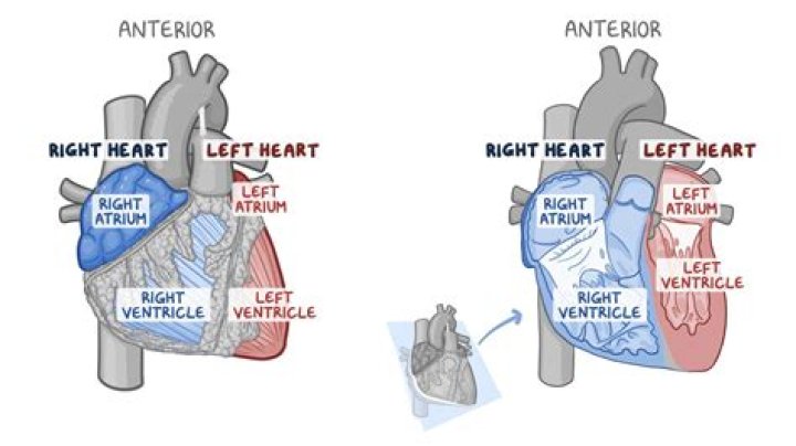
What are the 2 sides of the heart?
Your heart is divided into two separate pumping systems, the right side and the left side. The right side of your heart receives oxygen-poor blood from your veins and pumps it to your lungs, where it picks up oxygen and gets rid of carbon dioxide.