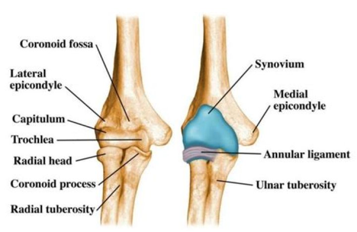
The trochlea has the capitulum
What is lateral Trochlear?
Lateral trochlear inclination (LTI) is a described measure of trochlear morphologic characteristics. 5. The LTI is measured on axial MRI sequences at the level of the most proximal extent of the trochlear cartilaginous surface.
What is the lateral femoral trochlea?
The subchondral bone of the trochlea is normally seen as a dense white line, which remains parallel to the subchondral bone of the lateral femoral condyle. The intersection of those two lines is indicative of severe trochlear dysplasia and has been named “the lateral trochlear sign.”
Where is the trochlea located in the knee?
The trochlea is a groove in the femur bone underneath the kneecap (patella). The walls of the trochlea stabilize the patella and allow it to glide down the center of the trochlea as the knee bends.Where is the trochlea of the femur?
The trochlea of femur (femoral trochlea) is the cranial cartilaginous part of distal femur, for articulation with the patella fo form the femoral patellar joint. It consists of a groove bounded by the medial and a lateral ridges.
Where is the Trochlear groove?
When the knee is bent, the undersurface of the kneecap (the patella) lies in an area known as the trochlear groove. The sides of the patella and the walls of the groove should be almost parallel. The normal shape of the trochlea groove is concave.
Where is the Trochlear groove located?
The trochlear notch (/ˈtrɒklɪər/), also known as semilunar notch and greater sigmoid cavity, is a large depression in the upper extremity of the ulna that fits the trochlea of the humerus (the bone directly above the ulna in the arm) as part of the elbow joint. It is formed by the olecranon and the coronoid process.
Can you move your kneecap side to side?
Injuries from sports, overuse, or trauma can cause the patella to move slightly off and not track properly in the trochlear groove. In most cases, the kneecap shifts to the outside of the leg, but it can also move towards the inside.Is the trochlea medial or lateral?
In the human arm, the humeral trochlea is the medial portion of the articular surface of the elbow joint which articulates with the trochlear notch on the ulna in the forearm.
What is the back of the knee called?The shallow depression formed at the back of the knee is called the popliteal fossa; it is formed at the junction of the femur and tibia. There is a muscle here on the floor of the popliteal fossa which is the deepest muscle of the knee joint.
Article first time published onDoes Trochlear dysplasia require surgery?
Knee Joint Trochlear dysplasia is a condition where the trochlear groove is abnormally shaped, causing the patella to slip out of the groove or dislocate. Trochleoplasty is a surgical procedure that reshapes the trochlea to prevent patellofemoral recurrent instability, and associated pain and disability.
What is the chondral?
Cartilage, or chondral, damage is known as a lesion and can range from a soft spot on the cartilage (Grade I lesion) or a small tear in the top layer to an extensive tear that extends all the way to the bone (Grade IV or “full-thickness” lesion).
Is Trochlear dysplasia congenital?
Trochlear dysplasia is characterized by abnormal trochlear morphology and a shallow groove. It is associated with recurrent patellar dislocation, but it is unclear whether the dysplasia is congenital, the result of lateral tracking and chronic instability, or caused by a combination of factors.
What is trochlea of Talus?
The trochlea of talus is a convex articular surface for the proximal part of articulation with tibia and fibula (only with tibia in horses). The trochlea presents a sagittal groove that articulates with the sagittal ridge of cochlea of tibia.
What does the trochlea of the femur articulate with?
The capitulum laterally articulates with the radius; the trochlea, a spool-shaped surface, articulates with the ulna.
What is shallow femoral trochlea?
Femoral trochlear dysplasia is characterized by an abnormally shallow trochlear groove. Disengagement of the patella from the shallow femoral trochlea is common in FTD and is a predisposing risk factor to recurrent patellar dislocation and subsequent premature osteoarthrosis.
Where is radial notch of ulna?
Anatomical terms of bone The radial notch of the ulna (lesser sigmoid cavity) is a narrow, oblong, articular depression on the lateral side of the coronoid process; it receives the circumferential articular surface of the head of the radius.
Where is the olecranon fossa located?
The olecranon fossa is located on the posterior surface of the distal humerus, where it receives the proximal ulna during full extension of the arm.
Where are the capitulum and trochlea located quizlet?
More distally, at the elbow, the capitulum of the humerus articulates with the head of the radius, and the trochlea of the humerus articulates with the trochlear notch of the ulna. is situated lateral to the head of the humerus and posterolateral to the lesser tubercle.
What does patellofemoral arthritis feel like?
Patients experiencing patellofemoral knee arthritis will have kneecap pain and stiffness and often swelling in the front part of the knee that typically worsens when walking on inclined terrain, going up and down stairs, squatting or rising from a seated position.
What is the best treatment for chondromalacia patella?
The most common way to treat symptoms of chondromalacia patella is to rest the knee. Other ways to treat the symptoms include: Placing of an ice or cold pack to the area for 15-20 minutes, four times daily, for several days. Do not apply ice directly to the skin.
Can your kneecap wear away?
The cartilage surfaces behind the kneecap are the thickest in the entire human body and usually begin to wear out after the age of 15. Almost all people have evidence of cartilage damage on kneecap cartilage when we perform arthroscopic surgery.
Where is the lateral epicondyle of the humerus?
There are bony bumps at the bottom of the humerus called epicondyles, where several muscles of the forearm begin their course. The bony bump on the outside (lateral side) of the elbow is called the lateral epicondyle.
Where is the lateral condyle of femur?
The lateral condyle is one of the two projections on the lower extremity of the femur. The other one is the medial condyle. The lateral condyle is the more prominent and is broader both in its front-to-back and transverse diameters.
What bones have a trochlea?
The trochlea is the roughly hourglass-shaped feature on the distal end of the humerus. It articulates with the trochlear notch of the ulna.
What is a floating knee?
Floating knee is a flail knee joint resulting from fractures of the shafts or adjacent metaphyses of the femur and ipsilateral tibia. [1, 2] Blake and McBryde initially described this injury, which is generally caused by high-energy trauma.
Why does the patella dislocate laterally?
Patellar dislocations tend to occur in a lateral direction, partly because the direction of pull of the quadriceps muscle is slightly lateral to the mechanical axis of the limb. Medial instability is rare and more likely to result from congenital conditions, quadriceps atrophy, or iatrogenically.
What dislocated knee feels like?
Symptoms of a dislocated kneecap a “popping” sensation. severe knee pain. being unable to straighten the knee. sudden swelling of the knee.
What causes pain on back of leg behind knee?
What is pain behind the knee? Some of the most common causes of pain behind the knee (posterior knee pain) include, Baker’s cyst, arthritis, infection, injury, tumor, or deep vein thrombosis. Since the knee is the largest and most complex joint in the body, it makes sense that it might hurt sometimes.
What is the side of your knee called?
The lateral side of the knee is the side that is away from the other knee. Structures on the medial side usually have medial as part of their name, such as the medial meniscus. The term anterior refers to the front of the knee, while the term posterior refers to the back of the knee.
What causes pain behind knee after sitting?
What Causes Pain Behind The Knee After Sitting? Pain behind the knee after sitting for prolonged periods is often caused by arthritis. When we sit still, the fluid that lubricates the knee joint dries out slightly so when we then stand up, there is less cushioning.